 |
 |
 |
|
||||||||||||||||||||||||
 | ||||||||||||||||||||||||
 | ||||||||||||||||||||||||
 | ||||||||||||||||||||||||
Confocal Microscopy Image Gallery
Rat Brain Tissue Sections
Hypothalamus
The hypothalamus is one of the main components of the diencephalon, which develops from the embryonic forebrain. The structure is linked ventrally to the thalamus, but is demarcated from that region by a shallow groove called the hypothalamic sulcus.

The floor and part of the walls of the third ventricle of the brain are formed by the hypothalamus, which is very small, only accounting for about 4 grams of the total weight of the human brain. Despite its diminutive size, many basic functions that are critical to survival are carried out by the hypothalamus. The functions of the structure are chiefly controlled by projection neurons located in specific nuclei or small groups of nuclei that interact with effectors in other regions of the central nervous system. Three mediolateral zones of these nuclei have been identified: the periventricular zone, the middle zone, and the lateral zone.
Input to the hypothalamus arrives via both circulatory and neural routes. Chemical, hormonal, and certain physical information, such as body temperature, are received by the hypothalamus from the blood as it circulates through the brain structure. The hypothalamus obtains other types of data from the medulla oblongata and the midbrain. Signals based on information acquired by receptors that comprise part of the autonomic nervous system are sent to the hypothalamus from projection neurons located in the nucleus solitarus of the medulla. Input regarding the state of mental arousal, however, is communicated by both the reticular formation and the monoaminergic nuclei located in the midbrain. Similar to hypothalamic input, output from the hypothalamus can be sent via the circulatory system or neural pathways.
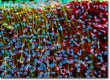
The rat hypothalamus coronal section presented above was immunofluorescently labeled for two different intermediate filament proteins, vimentin and glial fibrillary acidic protein. After the specimen was fixed, permeabilized, and blocked with 10-percent normal goat serum, it was treated with a cocktail of mouse anti-vimentin and rabbit anti-GFAP primary antibodies followed by goat anti-mouse and anti-rabbit secondary antibodies (IgG) conjugated to Alexa Fluor 488 and Alexa Fluor 568, respectively. The far-red fluorescent DNA probe DRAQ5 (pseudocolored cyan) was subsequently employed as a nuclear counterstain. Images were recorded with a 40x oil immersion objective using a zoom factor of 1.4 and sequential scanning with the 488-nanometer spectral line of an argon-ion laser, the 543-nanometer line from a green helium-neon laser, and the 633-nanometer line of a red helium-neon laser. During the processing stage, individual image channels were pseudocolored with RGB values corresponding to each of the fluorophore emission spectral profiles unless otherwise noted above.
Additional Confocal Images of Rat Hypothalamus Tissue Sections
GFAP and Vimentin Distribution in the Rat Hypothalamus - Glial fibrillary acidic protein is strongly and specifically expressed in astrocytes and certain other astroglia present in the brain. The protein was targeted in the section of rat hypothalamus featured in this confocal image with rabbit anti-GFAP primary antibodies followed by goat anti-rabbit secondary antibodies conjugated to Alexa Fluor 568. The intermediate filament protein vimentin was also targeted in the tissue section with a mouse anti-vimentin antibody detected with a goat anti-mouse secondary antibody conjugated to Alexa Fluor 488. Cell nuclei were counterstained with DRAQ5.
Detecting Neurofilaments and Intermediate Filaments in Coronal Brain Sections with Immunofluorescence - In a double immunofluorescence experiment, a coronal section of rat hypothalamus tissue was treated with a cocktail of mouse anti-vimentin (intermediate filaments) and chicken anti-NF-H (neurofilaments) primary monoclonal antibodies followed by goat anti-mouse and anti-chicken secondary antibodies (IgG) conjugated to Alexa Fluor 488 and Alexa Fluor 568, respectively. The specimen was counterstained for nuclei with DRAQ5, a far-red fluorescent DNA probe.
Rat Brain Hypothalamus Tissue Triple Stained with Alexa Fluor 488, Alexa Fluor 568, and DRAQ5 - The proximity between glial fibrillary acidic protein and vimentin, both of which are members of the class III intermediate filament protein family, was visualized in rat hypothalamus tissue presented in this section. Before being treated with a cocktail of rabbit anti-GFAP and mouse anti-vimentin primary antibodies, the specimen was fixed, permeabilized, and blocked with 10-percent normal goat serum. The primary targets were visualized with goat anti-rabbit and anti-mouse secondary antibodies conjugated to Alexa Fluor 568 and Alexa Fluor 488, respectively. In order to detect cell nuclei, the tissue section was counterstained with DRAQ5.
NF-H, Vimentin, and DNA in a Hypothalamus Tissue Section - Neurofilaments, which are intermediate filaments that are found in neuronal cells, were immunofluorescently labeled in the coronal rat brain section featured in this confocal image with a primary antibody directed against one of their protein subunits (NF-H) followed by a secondary antibody conjugated to Alexa Fluor 568. In addition, the intermediate filament protein vimentin was targeted with an anti-vimentin primary antibody visualized with a secondary antibody conjugated to Alexa Fluor 488. DRAQ5 served as a nuclear counterstain.
Class III Intermediate Filament Proteins Immunofluorescently Targeted with Antibodies Conjugated to Alexa Fluor Dyes - The intermediate filament protein vimentin is expressed by a number of different types of mesenchymal cells and in the precursor cells of astrocytes and neurons, while the closely related glial fibrillary acidic protein is specifically expressed in various astroglial cells and neural stem cells. These two proteins were visualized in a coronal rat brain section featuring the hypothalamus with mouse anti-vimentin and rabbit anti-GFAP primary antibodies detected with goat anti-mouse and anti-rabbit secondaries conjugated to Alexa Fluor probes (488 and 568, respectively). Nuclear DNA was labeled with the red-absorbing counterstain DRAQ5.
Visualizing GFAP and Vimentin in a Coronal Tissue Section of Rat Brain - A rat hypothalamus coronal tissue section was immunofluorescently labeled for the class III intermediate filament proteins vimentin and glial fibrillary acidic protein (GFAP). After the specimen was fixed, permeabilized, and blocked with 10-percent normal goat serum, it was treated with a cocktail of mouse anti-vimentin and rabbit anti-GFAP primary antibodies followed by goat anti-mouse and anti-rabbit secondary antibodies (IgG) conjugated to Alexa Fluor 488 and Alexa Fluor 568, respectively. In order to detect cell nuclei, the tissue section was subsequently treated with DRAQ5, a far-red fluorescent DNA probe.
Contributing Authors
Nathan S. Claxton, Shannon H. Neaves, and Michael W. Davidson - National High Magnetic Field Laboratory, 1800 East Paul Dirac Dr., The Florida State University, Tallahassee, Florida, 32310.
