Fluorescence Microscopy Digital Image Gallery
Specimens featured in the fluorescence digital image gallery are derived from a combination of stained thin sections, whole mounts, suspensions, smears, and several additional mounting techniques. Stained tissue culture cells and thin sections were labeled with either fluorescent dyes or common histology stains such as eosin, fast green, and safranin. Fluorescence microscopy and photomicrography was conducted by Charles D. Howard. Background research for the figure captions was provided by Elise Sessions. Kathleen Carr supervised the authoring and assembly of text for each of the gallery entries.
 Alfalfa Root
 American Dog Tick
 Ants
Ants Antelope Hair
 Basswood Stem
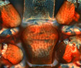 Bed Bugs
 Bird Lungs
 Bird Skin
 Black Grape Rot
 Cactus
 Corn Grain
 Corn Smut
 Dutchman's Pipe
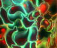 Elderberry
 Fava Bean Root Tip
 Female Pine Cones
 Fern Spores
 Fish Gill Filaments
 Fleas
Fleas Guinea Pig Hair
 Head Lice
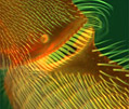 Honey Bee Leg
 Honey Bee Stinger
 House Fly Face
 Human Flea
 Hyaline Cartilage
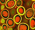 Human Roundworm
 Human Scalp
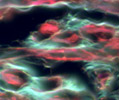 Human Spinal Cord
 Pony Belly Hair
 Kapok Fiber
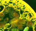 Leeches
 Lily Flower Bud
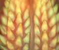 Lone Star Tick
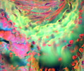 Compact Bones
 Spongy Bone
 Milkweed Fibers
 Mites
Mites Moss Tissue
 Mouse Intestines
 Mouse Kidney
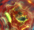 Mycorrhizal Fungi
 Obelia Hydroid
 Oleander Leaf
 Brown Rot of Peach
 Peach Leaf Curl
 Pine Root
 Pine Tree Pollen
 Pine Wood
 Privet Leaf
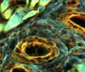 Raw Meat
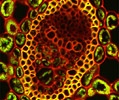 Rhizopus Rot
 Snail Radula
 Sweet Flag Grass
 Tilia
Tilia Trichina Worm
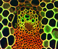 Wheat
Wheat Wheat Kernel
 Wheat Rust Pustule
