DIC Wavefront Relationships and Image Formation
The spatial relationship and phase difference between ordinary and extraordinary wavefronts in differential interference contrast (DIC) microscopy is a primary factor in determining how image formation occurs. This interactive tutorial explores wavefront relationships in the DIC microscope optical train, and how these relationships affect image formation.
The tutorial initializes with a typical DIC microscope optical train equipped with Wollaston prisms appearing in the window. A beam of linearly polarized light emerges from the polarizer and enters the lower Wollaston prism (labeled the Condenser Prism) before being separated into two orthogonal components and sheared at the quartz wedge interface. In the tutorial, the ordinary wavefront is represented by short red lines (parallel to the browser window) on a black ray trace, while the extraordinary wavefront is depicted by black dots (perpendicular to the browser window) on a red ray trace. The sheared orthogonal wavefronts are rendered parallel by the condenser and pass through the specimen before being collected by the objective and focused onto the interference plane of the second Wollaston prism (the Objective Prism). Recombined wavefronts emerge from the objective prism and pass through the analyzer to complete their journey through the virtual DIC microscope.
In order to operate the tutorial, use the Objective Prism Position slider to translate the objective prism laterally across the optical path of the microscope. As the prism is moved to the left or right, the Emerging Waveform (depicted in the upper right-hand corner of the tutorial window) changes from linear, through varying degrees of elliptical, and finally, to circular. When the waveform has some degree of elliptical or circular character, a portion of the light emerging from the objective prism can then pass through the analyzer to form an image. The Specimen Gradient slider can be utilized to increase the optical path gradient across the specimen, which will also produce a change in the waveform emerging from the objective prism. Wavefront relationships in both Nomarski and Wollaston prisms can be examined by toggling between the two prism types using the Objective Prism radio buttons (labeled Wollaston and Nomarski). The speed of wavefronts traveling through the virtual microscope can be increased or decreased using the Applet Speed slider.
After emerging from the condenser Wollaston or Nomarski prism at the aperture plane, the sheared ordinary and extraordinary coherent wavefronts are focused by the lens elements of the condenser and travel through the specimen before being collected by the objective. Along their trajectories between the condenser and objective, the wavefronts remain parallel to one another and are separated by a shear distance derived from the geometrical constraints of the condenser prism. The spatial separation between wavefronts (shear distance) varies with condenser and objective numerical aperture, but has practical limits between 0.1 and 1.5 micrometers, a linear range that is designed to be slightly smaller than (or in some cases equal to) the lateral resolution of the objective. Resolution in differential interference contrast can be increased (at the expense of contrast) by reducing the shear distance to about one-half the maximum resolution of the objective.
Most microscope manufacturers compromise on the shear distance versus resolution (and contrast) trade-off, and produce prisms that have a maximum shear distance of about 0.6 micrometers for lower magnification objectives (10x) down to a minimum approaching 0.15 micrometers for the higher magnification objectives (60x and 100x). Regardless of the shear distance, however, it is important to note that closely spaced wavefront pairs, spatially distributed across the entire microscope aperture, sample every point in the specimen to ultimately provide dual-beam interference at the image plane.

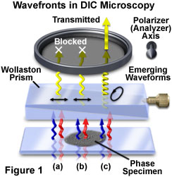
When undisturbed by the presence of a specimen, the coherent wavefront pairs experience identical optical path lengths between the specimen and image planes and arrive at the objective rear focal plane having the same phase relationship as when they left the condenser. The Nomarski prism located behind the objective recombines the wavefronts in the objective focal plane to generate linearly polarized light having an electric vector vibration orientation identical to that of the substage polarizer transmission axis. Linearly polarized wavefronts exiting the objective prism are blocked by the second polarizer (or analyzer), which has a transmission axis oriented perpendicular to that of the polarizer (Figure 1(a) and 1(b)). As a result, the image background observed in the viewfield appears very dark or black, a condition referred to as extinction.
Without specimen-induced phase shifts, the beamsplitting action of the condenser prism is exactly matched and reversed by the beam-recombining effect of the objective Nomarski prism to ultimately produce linearly-polarized light. In other words, when the microscope is properly configured for Köhler illumination (a critical prerequisite for high-resolution differential interference contrast microscopy), the condenser and objective operate in conjunction to project an image of the light source and condenser prism onto the objective prism. The objective Nomarski prism, which is inverted in orientation with respect to the condenser prism, introduces a phase shift that exactly compensates the linear phase shift between the wavefronts produced by the condenser prism. This action occurs for all paired wavefronts across the entire microscope aperture. The axes of both the condenser and objective prisms are parallel to each other and oriented at a 45-degree angle with respect to the transmission axes of the crossed polarizers (polarizer and analyzer). The orientation axis of the two prisms is termed the shear axis, an important concept that defines the axis of lateral separation between the ordinary and extraordinary wavefronts from the time they leave the condenser prism until they are recombined by the objective prism and arrive at the image plane.
In the event that coherent paired wavefronts encounter a phase gradient present in the specimen while passing from the condenser to the objective, wavefront distortion is induced and the waves will undergo a phase shift along the shear axis and traverse slightly different optical paths (although no change in polarization occurs). Upon arriving at the objective prism, the phase-shifted paired wavefronts are recombined to generate elliptically polarized light (in contrast to the linearly polarized light produced in the absence of a specimen). The electric vector of the resultant wavefront, which is no longer planar, sweeps out an elliptical pathway as it travels through the region between the objective prism and the analyzer (as illustrated in Figure 1(c)). Because a component of the elliptical wavefront is now parallel to the transmission axis of the analyzer, some portion of the wave will pass through the analyzer and produce plane-polarized light having a finite amplitude and ultimately being able to generate intensity in the image plane.
In summary, optical path gradients in the specimen induce phase shifts in the coherent paired wavefronts sheared by the condenser prism and passing through on parallel trajectories. These phase shifts are translated into phase differences by the objective Nomarski prism, creating elliptically polarized light that is capable of passing a linear component through the analyzer and creating an image. In fact, over the entire specimen field, the presence or absence of phase gradients creates a combination of linearly and elliptically polarized wavefronts that are selectively passed by the analyzer according to the azimuths of their vibrational planes. The wavefronts that are able to pass through the analyzer are all plane-parallel and can generate an amplitude image of the specimen through interference at the image plane. When the objective prism exactly compensates the effects of the condenser prism (as it does in Köhler illumination), the analyzer blocks wavefronts originating from all spatial locations of the field lacking phase shifts (no specimen phase gradients). The resulting background observed in the viewfield is dark (exhibiting total extinction) with the exception of regions displaying steep specimen refractive index or thickness gradients, which appear much brighter (usually in outline form). The perceived image appears very similar to images generated by the classical, but simple, darkfield illumination technique.
