
Deconvolution in Optical Microscopy
Section Overview:
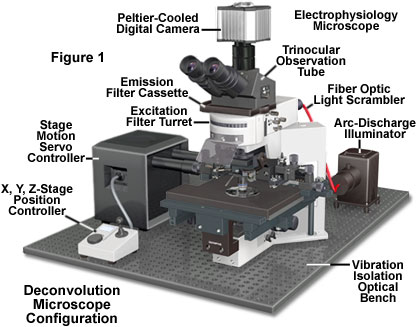
Deconvolution is a computationally intensive image processing technique that is being increasingly utilized for improving the contrast and resolution of digital images captured in the microscope. The foundations are based upon a suite of methods that are designed to remove or reverse the blurring present in microscope images induced by the limited aperture of the objective.
Practically any image acquired on a digital fluorescence microscope can be deconvolved, and several new applications are being developed that apply deconvolution techniques to transmitted light images collected under a variety of contrast enhancing strategies. One of the most suitable subjects for improvement by deconvolution are three-dimensional montages constructed from a series of optical sections.
Review Articles
-
Introduction to Deconvolution
Deconvolution is a computationally intensive image processing technique that is being increasingly utilized for improving the contrast and resolution of digital images captured in the microscope.
-
Algorithms for Deconvolution Microscopy
Discussed in this section are the most commonly utilized algorithms for deconvolution in optical microscopy can be divided into two classes: deblurring and image restoration.
-
Artifacts and Aberrations in Deconvolution Analysis
Artifacts are often not caused by computation, but by histology, optical misalignment, or electronic noise. To diagnose the source of an artifact, the first step is to compare the raw image with the deconvolved image.
-
Resolution Criteria for Deconvolution Analysis
Resolution in optical microscopy is often assessed by means of an optical unit termed the Rayleigh criterion, which was originally formulated for determining the resolution of two-dimensional telescope images, but has since spread into many other arenas in optics.
Selected Literature References and Internet Resources
-
Deconvolution Resources
Listed in this section are links to web resources on deconvolution analysis, including software packages, hardware (microscopes and accessories), and laboratories involved with the technology.
-
Selected Literature References
A number of review articles on deconvolution in optical microscopy have been published by researchers in the field and were utilized as references to prepare discussions included in this Digital Image Processing Section.