
Darkfield MIC-D Digital Image Gallery
Darkfield illumination transforms specimens into bright, highlighted structures superimposed on a very dark or black background. When the MIC-D digital microscope illuminator is positioned at highly oblique angles (over 25 degrees from the optical axis), semi-transparent specimens can be readily observed and captured with the accompanying interface software. This gallery demonstrates the darkfield imaging ability of the microscope on a wide variety of specimens.
 Acanthocephala
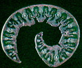 American Beachgrass
 Ants (Formicidae)
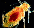 Aphids
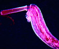
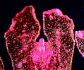 Aurelia
 Bacterial Capsules
 Bauxite Ore
 Butterfly Proboscis
 Canine Hookworm
 Chicken Embryos
 Cnidaria Fossil
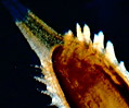 Dandelion Fruit
 Dead Leaf Butterfly
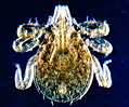 Deer Tick Larvae
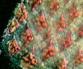 Dogfish Shark Scales
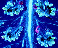 Fern Sori
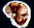 Foraminifera
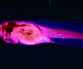 Frog Striated Muscle
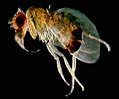 Fruit Fly
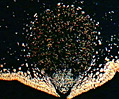 Fucus Conceptacle
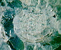 Goniatitic Cephalopod
 Grantia Sponge
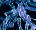 Grasshopper
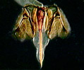 Honeybee Stinger
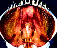 House Fly
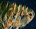 Human Flea
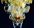 Human Head Louse
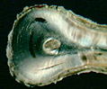 Human Tooth Root
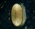 Insect Spiracles
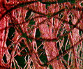 Jute
Jute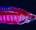 Lamprey Larva
 Leeches
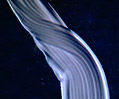 Lycra Spandex
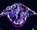 Metridium
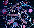 Mixed Green Algae
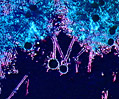 Mold Conidiophores
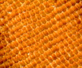 Monarch Butterfly
 Morpho Butterfly Wing
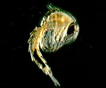 Mosquito Pupa
 Mucor Zygotes
 Parenchyma
 Pectinatella Bryozoans
 Pennaria Hydrozoa
 Pig Embryo
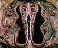 Pig Tooth
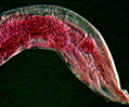 Pinworms
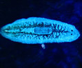 Planaria
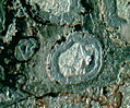 Pleistocene Bone
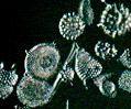 Radiolarians
 Sheep Ked Fly
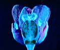 Shepherd's Purse
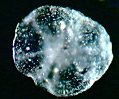 Sodium Chloride
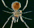 Spider
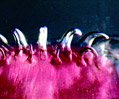 Tapeworm Scolex
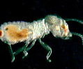 Termite
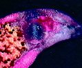 Trematode Flukes
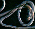 Trichuris Whipworms
 Varroa Mite
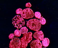 Volvox
 Zea (Corn) Kernel