
Brightfield Illumination MIC-D Digital Image Gallery
Featuring a wide spectrum of stained and unstained specimens, the MIC-D brightfield image gallery contains digital images that were captured using the microscope at a variety of zoom optical system magnifications. The images were corrected and adjusted with respect to contrast, brightness, sharpness, hue, color balance, and saturation using digital image processing tools available in the MIC-D software processing window.
 Acorn
Acorn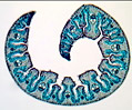 American Beachgrass
 Amphibian
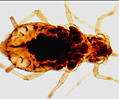 Aphid
Aphid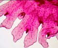 Aurelia Ephyra
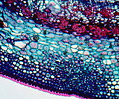 Basswood
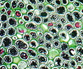 Bracken Fern
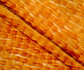 Butterfly Wing
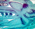 Cabbage Flower
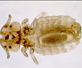 Canine Biting Louse
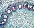 Corn Root Tissue
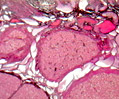 Corpus Luteum
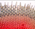 Ctenoid Fish
 Developing Long Bone
 Dicot Leaf Epidermis
 Dictydium cancellatum
 Dogfish Shark
 Druse Crystals
 Earthworm
 Epiglottis Elastic
 Adipose Tissue
 Fern Prothallium
 Fern Sporophyte
 Fetal Elastic
 Fetal Skull
 Filamentous Algae
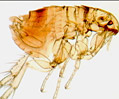 Flea
Flea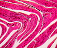 Frog Striated Muscle
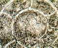 Goniatitic Cephalopod
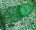
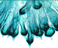 Codium
 Hairy Mammal
 Horsetail
 Auerbach's Plexus
 Human Bladder
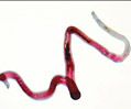 Human Blood
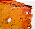 Human Compact Bone
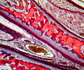 Human Fetus
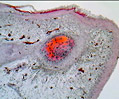 Human Fingernail
 Human Heart Tissue
 Lower Duodenum
 Human Lung Tissue
 Male Chromosomes
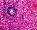 Human Spleen
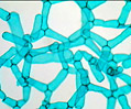 Hydrodictyon Algae
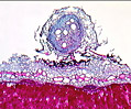 Legume Nodules
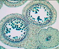 Lily Flower Bud
 Human Tongue
 Mammalian Elastic
 Mammalian Femur
 Mammalian Ganglion
 Taste Buds
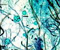 Mixed Green Algae
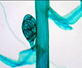 Moss Bulbils
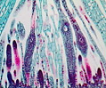 Moss Capsule
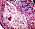 Mouse Tail
 Mucous Colon
 Nitelia Algae
 Obelia Hydroid
 Palm and Sole Skin
 Pennaria Hydrozoa
 Diatoms
 Peyer's Patches
 Pig Tooth Enamel
 Pine Mature Embryo
 Pine Needle Section
 Pine Stem Section
 Polysiphonia
 Potato Blight
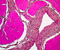 Primate Bladder
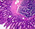 Primate Colon
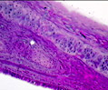 Primate Hyaline
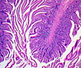 Primate Ileum
 Rhizopus Conjugation
 Scabies Infestation
 Shepherd's Purse
 Snake Cross-Section
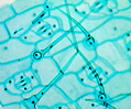 Spiderwort Leaf
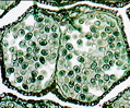 Staminate Pine Cone
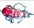 Starfish Larva
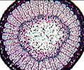 Sycamore Stem
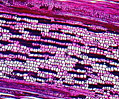 Sycamore Stem
 Tongue Muscle
 Trichinella Larvae
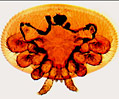 Varroa Mite
 Volvox
 Zea (Corn) Kernel
 Zygnema