 |
 |
 |
|
||||||||||||||||||||||||
 | ||||||||||||||||||||||||
 | ||||||||||||||||||||||||
 | ||||||||||||||||||||||||
Confocal Microscopy Image Gallery
Rat Tongue Taste Buds
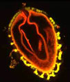
A thick section of rat tongue was triple stained with the nucleic acid fluorochrome DAPI, fluorescein isothiocyanate (FITC), and Texas Red to yield the striking confocal image presented below. Nuclei of the cells fluoresce a bright blue, while a high-affinity receptor for brain-derived neutrotrophic factor (TrkB) is stained green in the image. Protein gene products, distributed throughout the tissue, are stained red with the popular rhodamine derivative. The image was contributed by Shigeru Takami from the Department of Anatomy in the School of Health Science at Kyorin University, in Japan.

In mammals, the primary function of the tongue is to act as a mechanism for taste, although it is also important for human speech, suckling, and swallowing, as well as the grooming of some animals. The organ, which is a mass of striated muscle interspersed with glands and fat, is covered by a mucous membrane. It is along the top surface of this mucous membrane that the numerous projections called papillae and the clusters of cells called taste buds they contain are most heavily located. Each taste bud may hold as many as 75 taste receptors, but, classically, scientists have held that only four basic types of taste receptors exist, one for each taste sensation: salty, sweet, sour, and bitter. Modern research has, however, indicated that this traditional view is fundamentally flawed and that the taste buds and receptors are actually much more complex than formerly believed.
Confocal microscopy has frequently been utilized with great success during the contemporary reconsideration of the components and functions involved in the sensation of taste. The technique is particularly well suited for such research because it enables the examination of thick sections of tongue from a variety of animals. Also, when utilized with fluorescent markers, the approach facilitates the identification and understanding of minute structures within specimens, while optical sectioning followed by reconstruction processes results in accurate three-dimensional visualizations with amazing anatomical detail. Some of the questions that confocal microscopy is helping to address in the area include how information about taste quality is extracted by receptor cells, what electrophysiological activities take place during the process, and whether or not different morphological types of taste buds are truly representative of distinct cell types.
