 |
 |
 |
|
||||||||||||||||||||||||
 | ||||||||||||||||||||||||
 | ||||||||||||||||||||||||
 | ||||||||||||||||||||||||
Confocal Microscopy Image Gallery
Rat Brain Tissue Sections
Longitudinal Fissure
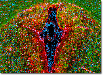
The longitudinal fissure is a deep grove in the brain that divides the right and left cerebral hemispheres from each other. Sometimes the cleft is alternatively referred to as the interhemispheric fissure.

The division of the cerebrum by the longitudinal fissure is incomplete. Located at the base of the cleft is a fibrous band of white matter called the corpus callosum that connects analogous regions on opposite sides of the brain. Communication between the hemispheres is carried out via the corpus callosum. The two halves of the brain also share a vasculature system, components of the system entering into the longitudinal fissure and interconnecting.
In human beings the cerebrum is the largest part of the brain, accounting for approximately two-thirds of the organís weight. The outer cortex of the human cerebrum is much more developed than it is in other animals. This difference is generally considered to be responsible for the greater cognitive capacity of humans, including the ability to perform abstract thought and produce complex speech. The cortex of the human brain exhibits numerous convolutions that allow the confined space of the skull to contain a greater cortical surface area. The elevated areas of the convolutions are called gyri and the grooves found between them are normally termed sulci. When a sulcus is especially deep, however, it is known as a fissure. The longitudinal fissure is the deepest fissure in the brain.
This horizontal thin section of rat brain clearly reveals the prominent longitudinal fissure. The specimen was fixed, permeabilized, blocked with 10-percent normal goat serum, and then treated with a cocktail of rabbit anti-GFAP (astroglia and neural stem cells) and chicken anti-NF-H (neurons) primary antibodies followed by goat anti-rabbit and anti-chicken secondary antibodies (IgG) conjugated to Alexa Fluor 568 and Alexa Fluor 488, respectively. Nuclei in the tissue section were subsequently targeted with DRAQ5 (pseudocolored cyan), a far-red fluorescent DNA probe. Images were recorded with a 20x objective using a zoom factor of 1.0 and sequential scanning with the 488-nanometer spectral line of an argon-ion laser, the 543-nanometer line from a green helium-neon laser, and the 633-nanometer line of a red helium-neon laser. During the processing stage, individual image channels were pseudocolored with RGB values corresponding to each of the fluorophore emission spectral profiles unless otherwise noted above.
Contributing Authors
Nathan S. Claxton, Shannon H. Neaves, and Michael W. Davidson - National High Magnetic Field Laboratory, 1800 East Paul Dirac Dr., The Florida State University, Tallahassee, Florida, 32310.
