 |
 |
 |
|
||||||||||||||||||||||||
 | ||||||||||||||||||||||||
 | ||||||||||||||||||||||||
 | ||||||||||||||||||||||||
Confocal Microscopy Image Gallery
Rat Brain Tissue Sections
Lateral Ventricle
The ventricular system is a network of interconnected cavities in the brain through which cerebrospinal fluid flows that functions in shock absorption, nutrient distribution, and detoxification. Two of these cavities, the lateral ventricles, have a complex C-shape that features several projections into different parts of the brain.

The anterior horn of each lateral ventricle projects into the frontal lobe of the brain, while the posterior and inferior horns respectively extend into the occipital and temporal lobes. The area of a lateral ventricle where the three horns meet is called the atrium. Most of the cerebrospinal fluid entering into the ventricular system is secreted by the choroid plexus in the walls of the lateral ventricles, but the choroid plexus of the narrow, slit-like third ventricle along the midline of the brain and the fourth ventricle positioned in the hindbrain exude the important substance as well, though in smaller amounts. Cerebrospinal fluid exits the ventricular system by means of three apertures in the fourth ventricle and is absorbed into the subarachnoid space.
If the ventricular system becomes obstructed, cerebrospinal fluid can build up in the ventricles, which become enlarged. This condition, called hydrocephalus, can result in a rapid increase in the head size of infants because the bones of their skulls are not fused together. As the pressure on the brain continues to increase, seizures and mental retardation may ensue. In older children and adults, the swelling of the ventricles cannot be accommodated by an expansion of the skull, so these patients often experience different symptoms, including headaches, vomiting, nausea, unsteadiness, blurred vision, personality changes, and impairment of mental functions. Hydrocephalus is almost always fatal if left untreated, but can often be remedied through the surgical insertion of a shunt to drain excess cerebrospinal fluid from the ventricles or a process called ventriculostomy that involves creating a small opening in the third ventricle of the brain.
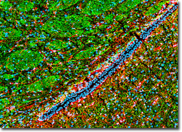
A lateral ventricle can be seen in the coronal thin section of rat brain presented in the digital image above, which was immunofluorescently labeled for heavy chain neurofilament subunits (neurons) and glial fibrillary acidic protein (astroglia and neural stem cells) with chicken anti-NF-H antibodies and rabbit anti-GFAP primary antibodies. The primary targets were visualized with goat anti-chicken and anti-rabbit secondary antibodies conjugated to Alexa Fluor 488 (green fluorescence emission) and Alexa Fluor 568 (red emission), respectively. Nuclear DNA was counterstained with DRAQ5 (pseudocolored cyan). Images were recorded with a 20x objective using a zoom factor of 1.3 and sequential scanning with the 488-nanometer spectral line of an argon-ion laser, the 543-nanometer line from a green helium-neon laser, and the 633-nanometer line of a red helium-neon laser. During the processing stage, individual image channels were pseudocolored with RGB values corresponding to each of the fluorophore emission spectral profiles unless otherwise noted above.
Contributing Authors
Nathan S. Claxton, Shannon H. Neaves, and Michael W. Davidson - National High Magnetic Field Laboratory, 1800 East Paul Dirac Dr., The Florida State University, Tallahassee, Florida, 32310.
