 |
 |
 |
|
||||||||||||||||||||||||
 | ||||||||||||||||||||||||
 | ||||||||||||||||||||||||
 | ||||||||||||||||||||||||
Confocal Microscopy Image Gallery
Rat Brain Tissue Sections
Cerebellum
The cerebellum is located posterior to the brain stem to which it is connected via bundles of nerves and is separated from the cerebral cortex by an extension of the dura mater called the tentorium cerebelli. Similar to the cerebrum, the cerebellum is divided into two hemispheres as well as an outer mantle of gray matter (termed the cerebellar cortex) and an internal core of white matter, which contains four bilaterally paired nuclei (fastigial, globose, emboliform, and dentate nuclei).

In humans the cerebellum comprises only about one-tenth of the total volume of the brain, but more than 50 percent of all its neurons are located in the structure. Cerebellar neurons are highly organized, and although various regions of the structure receive input from different sources and project to various motor systems, they appear to carry out comparable types of activity. This activity involves integrating and evaluating information regarding intent and action so that adjustments may be made in the motor systems to correct for any disparities between the two during the course of a movement.
The cortex of the cerebellum exhibits three layers, which contain a total of five different types of neurons. Four of these nerve cell types, including Purkinje, Golgi, stellate, and basket cells, are inhibitory, while the fifth variety, the granule cells, are excitatory. The central layer of the cerebellar cortex is the Purkinje cell layer, which is composed of a single stratum of Purkinje cell bodies. The axons of the Purkinje cells extend into the white matter that underlies the cortex, whereas their branching dendrites extend to the outermost layer of the cerebellar cortex, called the molecular layer. Stellate and basket cells are found in the molecular layer amidst the Purkinje dendrites and the axons of granule cells, the cell bodies of which are found in the innermost cortical stratum, the granular layer. Granule cell axons in the molecular layer are arranged parallel to the long axes of convolutions called folia and are consequently termed parallel fibers. The Purkinje dendrites run perpendicular to the parallel fibers.
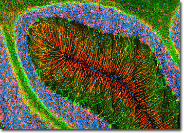
The neuron-specific class III beta-tubulin isoform was targeted in the sagittal thin section of rat cerebellum presented in the confocal image above with rabbit anti-beta-III-tubulin antibodies visualized with goat anti-rabbit secondary antibodies conjugated to Alexa Fluor 488 (green emission). In addition, glial fibrillary acidic protein (GFAP), a type III intermediate filament protein that is a primary structural element of astrocytes and neural stem cells, was immunofluorescently labeled with primary mouse anti-GFAP antibodies followed by goat anti-mouse Fab fragments conjugated to Alexa Fluor 568 (red emission). The specimen was counterstained for cell nuclei with DRAQ5 (pseudocolored cyan). Images were recorded with a 10x objective using a zoom factor of 1.4 and sequential scanning with the 488-nanometer spectral line of an argon-ion laser, the 543-nanometer line from a green helium-neon laser, and the 633-nanometer line of a red helium-neon laser. During the processing stage, individual image channels were pseudocolored with RGB values corresponding to each of the fluorophore emission spectral profiles unless otherwise noted above.
Additional Confocal Images of Rat Cerebellum Tissue Sections
Targeting GFAP and Class III beta-Tubulin in the Rat Cerebellum with Immunofluorescence - In a double immunofluorescence experiment, the rat cerebellum sagittal section presented in this section was fixed, permeabilized, blocked with 10-percent normal goat serum, and then treated with a cocktail of mouse anti-GFAP (glial fibrillary acidic protein) and rabbit anti-beta-III-tubulin (specific to neurons) primary antibodies followed by goat anti-mouse and anti-rabbit secondary antibodies (IgG) conjugated to Alexa Fluor 568 and Alexa Fluor 488, respectively. Nuclei in the specimen were subsequently targeted with DRAQ5.
Distribution of Neurons, Astroglia, and DNA in a Coronal Rat Brain Tissue Section - A coronal thin section of rat cerebellum was immunofluorescently labeled for heavy chain neurofilament subunits with chicken anti-NF-H antibodies followed by goat anti-chicken secondary antibodies conjugated to Alexa Fluor 488. Neurofilaments are specialized intermediate filaments solely found in neurons, especially in their axons. In addition, glial fibrillary acidic protein, which is expressed in various astroglia and neural stem cells, was targeted with rabbit anti-GFAP antibodies visualized with goat anti-rabbit secondary antibodies conjugated to Alexa Fluor 568. Cell nuclei were counterstained with DRAQ5, a far-red fluorescent DNA probe.
Horizontal Cerebellum Section Stained for GFAP and Cell Nuclei with Alexa Fluor 568 and DRAQ5 - Glial fibrillary acidic protein (GFAP), which is strongly and specifically expressed in astrocytes and certain other types of astroglia, was targeted in a horizontal thin section of rat cerebellum with rabbit anti-GFAP monoclonal antibodies followed by goat anti-rabbit secondary antibodies conjugated to Alexa Fluor 568. Cell nuclei were subsequently targeted in the specimen with DRAQ5.
Glial Fibrillary Acidic Protein and Heavy Chain Neurofilament Subunits in Rat Brain Tissue - Immunofluorescence was utilized to label neurofilaments and astrocytes in this thin section of rat cerebellum tissue. First, the specimen was fixed, permeabilized, blocked with 10-percent normal goat serum, and treated with a cocktail of chicken anti-NF-H and rabbit anti-GFAP primary antibodies. Then, to visualize the primary targets, the cerebellum section was treated with goat anti-chicken and anti-rabbit secondary antibodies (IgG) conjugated to Alexa Fluor 488 and Alexa Fluor 568, respectively. Finally, DRAQ5 was employed to counterstain cell nuclei.
Immunofluorescently Visualizing the Strata of the Rat Cerebellum - The high level of organization exhibited in the cerebellum is revealed in the sagittal rat brain section featured in this section. Note, nuclei, neural axons, and astrocytes chiefly appear in distinct layers. The cerebellar pattern was visualized by immunofluorescently labeling the specimen with rabbit anti-GFAP antibodies (glial fibrillary acidic protein; astrocytes and certain other stellate cells) and chicken anti-NF-H antibodies (neurofilaments; most abundant in axons of large projections neurons) followed by goat anti-rabbit and anti-chicken antibodies conjugated to Alexa Fluor 568 and Alexa Fluor 488, respectively. Cell nuclei were envisaged with the red-absorbing probe DRAQ5.
Cerebellar Tissue Triple Stained with Alexa Fluor 488, Alexa Fluor 568, and DRAQ5 - A rat cerebellum thin section was immunofluorescently labeled for heavy chain neurofilament subunits with chicken anti-NF-H monoclonal antibodies followed by goat anti-chicken secondary antibodies conjugated to Alexa Fluor 488. In addition, glial fibrillary acidic protein was targeted in the tissue section with rabbit anti-GFAP antibodies visualized with goat anti-rabbit secondaries conjugated to Alexa Fluor 568. A far-red fluorescent probe, DRAQ5, was used to counterstain cell nuclei.
Distribution of Astrocytes and Neurons in the Cerebellum - The coronal section of rat cerebellum presented in this confocal image was stained for astrocytes, neurons, and cell nuclei. Primary rabbit antibodies against glial fibrillary acidic protein, which is a member of the class III intermediate filament protein family specifically expressed in astrocytes and certain other stellate cells in the central nervous system, were visualized with goat anti-rabbit secondary antibodies conjugated to Alexa Fluor 568. Chicken antibodies against heavy chain neurofilament subunits, which are solely expressed in neurons, were simultaneously employed to target neural cells, and these primary targets were visualized with a secondary antibody (goat anti-chicken) conjugated to Alexa Fluor 488. To label cell nuclei, the specimen was subsequently treated with DRAQ5.
Imaging Glial Fibrillary Acidic Protein and Neurofilaments in Rat Brain Tissue Sections - Glial fibrillary acidic protein (GFAP) and heavy chain neurofilament proteins were targeted in a thin section of rat cerebellum with rabbit anti-GFAP and chicken anti-NF-H monoclonal antibodies followed by goat anti-rabbit and anti-chicken secondary antibodies conjugated to Alexa Fluor 568 and Alexa Fluor 488, respectively. The nuclear counterstain DRAQ5 (pseudocolored cyan) was subsequently employed in order to label cell nuclei.
Cerebellar Tissue Labeled for NF-H, GFAP, and DNA - Neurofilaments are specialized intermediate filaments only present in neurons that are usually composed of a combination of three proteins, known as NF-L, NF-M, and NF-H. These filaments were immunofluorescently labeled in this thin section of rat cerebellar tissue with chicken anti-NF-H antibodies followed by goat anti-chicken secondary antibodies conjugated to Alexa Fluor 488. In addition, glial fibrillary acidic protein, which is an intermediate filament protein found in the central nervous system in certain types of astroglia, was targeted with rabbit anti-GFAP antibodies visualized with secondary antibodies (goat anti-rabbit) conjugated to Alexa Fluor 568. Cell nuclei were counterstained with DRAQ5.
High Magnification Image of Rat Brain Coronal Section - This high magnification confocal image of a rat cerebellum coronal section was produced by probing the specimen with DRAQ5, Alexa Fluor 488, and Alexa Fluor 568. The two Alexa Fluor dyes were conjugated to secondary antibodies directed against primary chicken anti-NF-H antibodies and rabbit anti-GFAP antibodies in order to label heavy chain neurofilament subunits in neurons (Alexa Fluor 488; green emission) and glial fibrillary acidic protein in astrocytes and certain other astroglia (Alexa Fluor 568; red emission).
Detecting the Organization of Neurons and Astrocytes in a Cerebellum Section with Immunofluorescence - The cerebellum is a highly organized brain structure, as demonstrated by the image of a rat brain coronal section presented above. The specimen was immunofluorescently labeled for astrocytes and neurons with rabbit anti-GFAP monoclonal antibodies and chicken anti-NF-H antibodies followed by goat anti-rabbit and anti-chicken secondary antibodies conjugated to Alexa Fluor 568 and Alexa Fluor 488, respectively. GFAP is strongly and specifically expressed by various astroglia, and NF-H is solely found in the neurofilaments of neuronal cells. Cell nuclei were visualized with the red-absorbing probe DRAQ5.
Contributing Authors
Nathan S. Claxton, Shannon H. Neaves, and Michael W. Davidson - National High Magnetic Field Laboratory, 1800 East Paul Dirac Dr., The Florida State University, Tallahassee, Florida, 32310.
