Confocal Microscopy Image Gallery
Rat Brain Tissue Sections
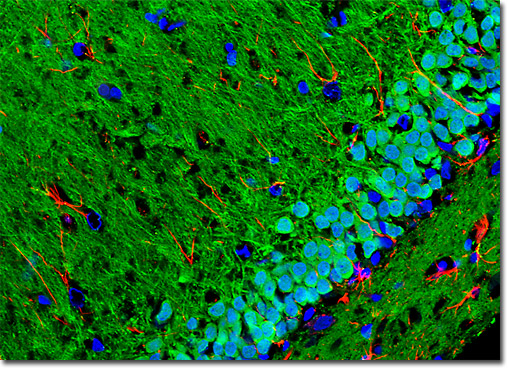
Amygdala
|
The calcium-binding protein calbindin and glial fibrillary acidic protein, a type III intermediate filament protein, were immunofluorescently labeled in the coronal rat amygdala tissue section presented above by treating the specimen with a cocktail of rabbit anti-calbindin and mouse anti-GFAP primary antibodies followed by goat anti-rabbit and anti-mouse secondary antibodies conjugated to Alexa Fluor 488 (green fluorescence) and Alexa Fluor 568 (red fluorescence), respectively. DRAQ5, a DNA-interactive agent that exhibits preferential intercalation at AT base pairs, was utilized to target cell nuclei. Images were recorded with a 40x oil immersion objective using a zoom factor of 1.6 and sequential scanning with the 488-nanometer spectral line of an argon-ion laser, the 543-nanometer line from a green helium-neon laser, and the 633-nanometer line of a red helium-neon laser. During the processing stage, individual image channels were pseudocolored with RGB values corresponding to each of the fluorophore emission spectral profiles, with the exception of DRAQ5, which was pseudocolored blue. View a larger version of this digital image. |
 |
 |
 |
 |
 |
 |
 |