 |
 |
 |
|
||||||||||||||||||||||||
 | ||||||||||||||||||||||||
 | ||||||||||||||||||||||||
 | ||||||||||||||||||||||||
Confocal Microscopy Image Gallery
Rat Brain Tissue Sections
Amygdala
The amygdala is a subcortical nuclear complex that plays a critical role in the coordination of emotional states and their physical expression. It is considered part of the limbic system and is located near the base of the cerebral hemispheres.

Numerous direct connections between the amygdala, the hippocampal formation, and neocortical areas exist that are extremely important for the functioning of the amygdala, which must receive information about the emotions associated with particular stimuli in order to influence behavior. Studies indicate that the amygdala is particularly involved with the emotional experience of fear and anxiety, which can be produced in humans when this region of the brain is electrically stimulated. Experimental investigations with laboratory animals have also demonstrated that impairment of the amygdala results in placidity and tameness.
Three divisions of the amygdala are often recognized: the basolateral nuclear group, the centromedial group, and the cortical nucleus. The most differentiated part of the amygdala is the basolateral nuclear group, which consists of the basal nucleus, the accessory basal nucleus, and the lateral nucleus. This collection of nuclei serves as the recipient of afferent sensory information to the amygdala and is thought to be chiefly responsible for the integration of this input. Some researchers have produced evidence that the basolateral nuclei may be involved with linking locational cues to particular rewards (context conditioning). The major region of output from the amygdala is the central nucleus, which is usually considered part of the centromedial nuclear group.
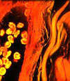
The calcium-binding protein calbindin and glial fibrillary acidic protein, a type III intermediate filament protein, were immunofluorescently labeled in the coronal rat amygdala tissue section presented above by treating the specimen with a cocktail of rabbit anti-calbindin and mouse anti-GFAP primary antibodies followed by goat anti-rabbit and anti-mouse secondary antibodies conjugated to Alexa Fluor 488 (green fluorescence) and Alexa Fluor 568 (red fluorescence), respectively. DRAQ5, a DNA-interactive agent that exhibits preferential intercalation at AT base pairs, was utilized to target cell nuclei. Images were recorded with a 40x oil immersion objective using a zoom factor of 1.6 and sequential scanning with the 488-nanometer spectral line of an argon-ion laser, the 543-nanometer line from a green helium-neon laser, and the 633-nanometer line of a red helium-neon laser. During the processing stage, individual image channels were pseudocolored with RGB values corresponding to each of the fluorophore emission spectral profiles, with the exception of DRAQ5, which was pseudocolored blue.
Contributing Authors
Nathan S. Claxton, Shannon H. Neaves, and Michael W. Davidson - National High Magnetic Field Laboratory, 1800 East Paul Dirac Dr., The Florida State University, Tallahassee, Florida, 32310.
