 |
 |
 |
|
||||||||||||||||||||||||
 | ||||||||||||||||||||||||
 | ||||||||||||||||||||||||
 | ||||||||||||||||||||||||
Applications in Confocal Microscopy
Fluorescence Photobleaching Investigations
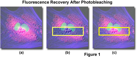
Both fluorescence loss in photobleaching (FLIP) and the related methodology of recovery after photobleaching (FRAP) are techniques for observing the movement of intracellular materials through photobleaching of fluorescence. A specific area of a floating fluorescent dye on a cell membrane, an organelle (endoplasmic reticulum and Golgi apparatus) membrane, or a floating fluorescence-labeled protein on these membranes is bleached, and the loss or recovery of fluorescence is observed to examine fluidity in the lateral direction (see Figure 1). The techniques of FLIP and FRAP are also used to confirm the continuity of membranes.

Recently, a study was conducted in which cytoskeletal proteins were labeled with fluorescent dye to confirm how turnover (replacement) of proteins constituting microtubules and intermediate-size filaments (10-nanometer filaments) occurs (Vikstrom et al., 1992). The techniques presented in this study can be used to determine whether the turnover occurs at the end or in the middle of the filaments.
Experiments using FLIP and FRAP have recently become much easier to carry out due to the use of green fluorescent proteins (GFPs). GFPs can be expressed in cells as a fusion protein with the target protein. Before GFPs became available, the observation of protein behavior in the cytoskeleton required proteins to be purified, fluorescently-labeled and introduced into the cell by means of microinjection.
Fluorescence Loss in Photobleaching
The continuity of organelle membranes in the cell including the endoplasmic reticulum, the Golgi apparatus and the nucleus has been verified by FLIP. After the area of interest has been photobleached, fluorescence is rapidly recovered by means of diffusion. When the same area has been bleached repeatedly, the fluorescence of the whole organelle membrane is lost, indicating that the fluorescence is floating on the organelle membrane.
Fluorescence Recovery After Photobleaching
A fluorescent dye previously conjugated with the target protein molecules or lipid membrane is bleached at the area of interest in the specimen and FRAP is used to observe how the fluorescence in the bleached portion recovers through dye turnover by means of diffusion.
Internet Resources
- Fluorescence Recovery After Photobleaching - In an experiment conducted by Dr. A. Malcolm Campbell (Molecular Biology, Department of Biology, Davidson College, Davidson, NC, USA), fluorescent dye previously conjugated with the target molecules was bleached at the area of interest in the specimen. The technique can also be used to determine if a protein is able to move within a membrane (high percent recovery with a fast mobility), or whether it is tethered other structural components of the cell (low percent recovery with a slow mobility).
Bibliography
Vikstrom, K. L. et al., Journal of Cell Biology, 118: 121-129 (1992).
