 |
 |
 |
|
||||||||||||||||||||||||
 | ||||||||||||||||||||||||
 | ||||||||||||||||||||||||
 | ||||||||||||||||||||||||
Three-Dimensional Imaging
The Olympus FluoViewTM confocal microscopes can be utilized to collect multiple optical sections (image stacks) along the specimen z-axis in order to obtain accurate three-dimensional structural analysis.
The Olympus FluoViewTM microscope software is capable of generating composite and multi-dimensional views of optical section data acquired from z-series image stacks. The three-dimensional rendering features of the software package can be employed to create either a single three-dimensional representation of the specimen or a video (movie) sequence compiled from different views of the specimen volume. These sequences often mimic the effect of rotation or similar spatial transformation that enhances the appreciation of the specimen's three-dimensional character. In addition, the software enables investigators to conduct measurements of length, volume, and depth, and specific parameters of the images, such as opacity, can be interactively altered to reveal internal structures of interest at differing levels within the specimen.
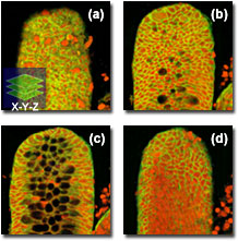
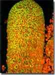
A typical stack of optical sections (often referred to as a z-series) through a human colon crypt revealing internal variations in fluorescence emission wavelengths is illustrated in the figure on the right. Optical sections were gathered in 0.5-micrometer steps perpendicular to the z-axis (microscope optical axis) using a dual argon-ion (488 nanometer; green fluorescence) and green helium-neon (543 nanometer; red fluorescence) laser system.
One of the foremost advantages of laser scanning confocal microscopy is the ability to serially produce thin (0.5 to 1.5 micrometers) optical sections through fluorescent specimens that have a thickness ranging up to 50 micrometers or more. The image series is collected by coordinating incremental changes in the microscope fine focus mechanism (using a stepper motor) with sequential image acquisition at each step. With the FluoViewTM software, the upper and lower limit of z scanning can be specified interactively by actually scanning the specimen or by direct input of the numerical value (the software settings panel is illustrated below).
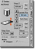
Image information is restricted to a well-defined plane, rather than being complicated by signals arising from remote locations in the specimen. Contrast and definition are dramatically improved over widefield techniques due to the reduction in background fluorescence and improved signal-to-noise. Furthermore, optical sectioning eliminates artifacts that occur during physical sectioning and fluorescent staining of tissue specimens for traditional forms of microscopy. The non-invasive confocal optical sectioning technique enables the examination of both living and fixed specimens under a variety of conditions with enhanced clarity.
The FluoViewTM three-dimensional imaging software can acquire x-y-z and display x-y cross sectional images quickly and continuously in increments of 0.01 micrometers due to the precision driving mechanism that enables tight step control. This feature is available on FluoViewTM scan heads attached to Olympus BX61, BX61WI, and IX81 microscopes, which are capable of generating high quality continuous cross sectional images. The three-dimensional function also provides extended focusing of images, stereo views, and animations to help explore the complete three-dimensional volume structure of the specimen.

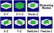
Optical sections are not restricted to the perpendicular lateral (x-y) plane with the FluoViewTM software, but can also be collected and displayed in transverse planes. Vertical sections in the x-z and y-z planes (parallel to the microscope optical axis, see the figure of the colon crypt on the left) can be readily generated. Thus, the specimen appears as if it had been sectioned in a plane that is perpendicular to the lateral axis. In practice, vertical sections are obtained by combining a series of x-y scans taken along the z axis with the software, and then projecting a view of fluorescence intensity as it would appear should the microscope hardware have been capable of physically performing a vertical section.