 |
 |
 |
|
||||||||||||||||||||||||
 | ||||||||||||||||||||||||
 | ||||||||||||||||||||||||
 | ||||||||||||||||||||||||

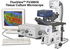
The Olympus FV300 is a point-scanning, point-detection, confocal laser scanning microscope designed for biology research applications. Excellent resolution, efficiency of excitation, intuitive user interface and affordability are fundamental characteristics of the FV300. The system permits simultaneous collection of up to 3 detection channels and may be configured on either the IX2 inverted research microscope platform or the BX2 upright research microscope platform.
The FluoViewTM FV300 scanning unit combines maximum optical efficiency with easy, one-touch selection of pinholes and filters under manual control. The system corrects aberrations ranging from the visible to near-infrared wavelengths, allowing aberration-free imaging for a wide range of applications.

Included in the compactly-designed scanning unit are a fiber optic coupler for incoming laser light sources, a beam collimator, laser adjustment neutral density filter turret, dichromatic mirrors, polarizer, x-y galvanometer-driven scan mirrors, pinhole turret, photomultipliers, beamsplitters, fluorescence filters, and a collector lens.
Superior Flexibility
Empty filter holders are available for the FV300 scanning unit as an option to allow operators the ability to design custom dichromatic mirrors, emission beamsplitters, and barrier filters for specific applications.
Two high-sensitivity photomultiplier tubes (PMTs) are located directly within the FV300 confocal fluorescence emission light path in the scanning unit for high sensitivity detection of the fluorescence signal. A separate, dedicated PMT may be used for the simultaneous detection of high-resolution brightfield or differential interference contrast (DIC) images with which the confocal fluorescence images may be overlaid.
Up to three channels of detection may be imaged simultaneously. Images may be scanned in any pixel array size up to 2048 x 2048, with each image digitized to 4096 gray levels (12-bit), permitting observation of fine image detail. Images may also be acquired in an automated sequential mode in order to reduce spectral crosstalk between channels for multi-color images.
System Compatibility
The FV300 scanning unit can be coupled with upright and inverted microscopes, including the BX51, BX51WI, BX61, BX61WI, and AX80 series of upright microscopes and the IX71 and IX81 inverted microscopes.
Features of the scanning unit include up to three imaging channels for ultraviolet, visible, and infrared lasers (into a single laser port) with a selection of five pinhole apertures. Laser control is provided by a laser combiner coupled to software on the host computer. The software interface is user-friendly and intuitive.
