 |
 |
 |
|
||||||||||||||||||||||||
 | ||||||||||||||||||||||||
 | ||||||||||||||||||||||||
 | ||||||||||||||||||||||||
Confocal Microscopy Image Gallery
Rat Brain Tissue Sections
Thalamus
The thalamus is the largest part of the diencephalon, which is located between the brain stem and the cerebral hemispheres. In human adults, the thalamus is about the size of a small egg. The structure usually appears as a double-lobed mass of gray matter positioned superior to the hypothalamus, from which its division is marked by a small groove termed the hypothalamic sulcus.

Together the thalamus and hypothalamus form the lateral wall of the third ventricle. In most humans, the two lobes of the thalamus are connected across the narrow ventricle by a nuclear mass called the interthalamic adhesion or massa intermedia, though in about 20 percent of the population the thalami are structurally separated from each other. A small bundle of nerve fibers connected to the limbic system, known as the stria medullaris, runs along the dorsomedial margin of the thalamus and another bundle of fibers called the stria terminalis is located at the boundary between the thalamus and the caudate nucleus, which is an important region of the basal ganglia.
Unlike other regions of the diencephalon, the thalamus does not appear to play a notable role in cyclical behaviors. Instead, the thalamus functions in motor control by being the primary recipient of motor information transmitted by the cerebrum. The thalamus is also a receiver of all auditory, somatosensory, and visual sensory signals from diverse areas of the brain, acting as the final gateway impulses must pass on their way to higher brain centers where additional interpretation and integration take place. Numerous groups of specialized neurons, termed nuclei, are present in the thalamus, which is divided into three primary nuclear masses (anterior, medial, and lateral) by a roughly Y-shaped stratum of nerve fibers called the internal medullary lamina. Within the internal medullary lamina there are embedded several intralaminar nuclei, and lateral to the primary masses of nuclei is a layer of nerve fibers called the lateral medullary lamina. Directly beneath the lateral medullary lamina is a sheet-like collection of neurons that comprise the reticular nucleus.
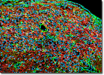
Immunofluorescence was utilized to label the neurofilaments of neurons and glial fibrillary acidic protein (GFAP) present in various astroglia in a coronal section of rat thalamus tissue. First, the specimen was fixed, permeabilized, blocked with 10-percent normal goat serum, and treated with a cocktail of chicken anti-NF-H (heavy chain neurofilament subunits) and rabbit anti-GFAP primary antibodies. Then, to visualize the primary targets, the tissue section was treated with goat anti-chicken and anti-rabbit secondary antibodies (IgG) conjugated to Alexa Fluor 568 and Alexa Fluor 488, respectively. Finally, DRAQ5 (pseudocolored cyan) was employed to counterstain cell nuclei. Images were recorded with a 20x objective using a zoom factor of 1.5 and sequential scanning with the 488-nanometer spectral line of an argon-ion laser, the 543-nanometer line from a green helium-neon laser, and the 633-nanometer line of a red helium-neon laser. During the processing stage, individual image channels were pseudocolored with RGB values corresponding to each of the fluorophore emission spectral profiles unless otherwise noted above.
Contributing Authors
Nathan S. Claxton, Shannon H. Neaves, and Michael W. Davidson - National High Magnetic Field Laboratory, 1800 East Paul Dirac Dr., The Florida State University, Tallahassee, Florida, 32310.
