 |
 |
 |
|
||||||||||||||||||||||||
 | ||||||||||||||||||||||||
 | ||||||||||||||||||||||||
 | ||||||||||||||||||||||||
Confocal Microscopy Image Gallery
Rat Brain Tissue Sections
Striatum
The basal ganglia are an interconnected group of subcortical neurons (termed nuclei) that are located deep in the interior of the cerebral hemispheres and are functionally involved in movement control, emotions, and cognition. The basal ganglia chiefly receive signals from the thalamus and the cerebral cortex and primarily pass information on to the brain stem and back to the thalamus, which returns the signals to the cortex.

The primary nuclei comprising the basal ganglia include the globus pallidus, the substantia nigra, the subthalamic nucleus, and the striatum. Most signals sent to the basal ganglia are received by the striatum, which is separated into three major divisions: the caudate nucleus, the putamen, and the nucleus accumbens. The axons of the neurons located in the striatum extend to both the globus pallidus and substantia nigra, which are the nuclei chiefly responsible for sending the output of the basal ganglia to other regions of the brain.
The vast majority of cells in the striatum are medium-spiny projection neurons, and some simple staining techniques seem to suggest that the structure is homogenous. Yet, in reality, the striatum is heterogenous in both composition and function. Neurotransmitters and neuromodulators are not evenly distributed throughout the striatum, but are concentrated in patches. These patches, often termed striosomes, are interspersed in a matrix of material that is histochemically distinct from them. Moreover, the neurons in the striosomes project to different areas than the cells located in the matrix, so their signals are targeted at different recipients. The full import of the compartmentalization of the striatum, however, has yet to be realized.
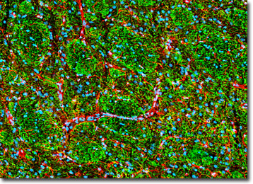
A coronal section of rat brain striatum (presented above) was immunofluorescently labeled for heavy chain neurofilament subunits with chicken anti-NF-H monoclonal antibodies followed by goat anti-chicken secondary antibodies conjugated to Alexa Fluor 488 (green). Neurofilaments, which are found in neurons, especially in their axons, are composed of light (NF-L) and medium (NF-M) chain subunits in addition to NF-H. Glial fibrillary acidic protein was also targeted in the tissue section by treating the specimen with rabbit anti-GFAP antibodies visualized with goat anti-rabbit secondary antibodies conjugated to Alexa Fluor 568 (red). Nuclear DNA was counterstained with DRAQ5 (pseudocolored cyan). Images were recorded with a 20x objective using a zoom factor of 1.3 and sequential scanning with the 488-nanometer spectral line of an argon-ion laser, the 543-nanometer line from a green helium-neon laser, and the 633-nanometer line of a red helium-neon laser. During the processing stage, individual image channels were pseudocolored with RGB values corresponding to each of the fluorophore emission spectral profiles unless otherwise noted above.
Contributing Authors
Nathan S. Claxton, Shannon H. Neaves, and Michael W. Davidson - National High Magnetic Field Laboratory, 1800 East Paul Dirac Dr., The Florida State University, Tallahassee, Florida, 32310.
