 |
 |
 |
|
||||||||||||||||||||||||
 | ||||||||||||||||||||||||
 | ||||||||||||||||||||||||
 | ||||||||||||||||||||||||
Confocal Microscopy Image Gallery
Rat Brain Tissue Sections
Pons
The brain stem is the lower portion of the brain that is continuous with the spinal cord. Three components comprise the brain stem, with the pons lying inferior to the midbrain and superior to the medulla oblongata. The posterior margin of the pons is divided from the cerebellum by the fourth ventricle and the aqueduct of Sylvius.

The pontine arteries that branch from the basilar artery, as well as the anterior inferior and superior cerebellar arteries, maintain a constant supply of blood to the pons, and masses of nerve fibers extending from the cerebral cortex synapse on the base of the structure at the pontine nuclei. Most of the input received at the nuclei is relayed from the motor and somatosensory regions of the cortex, and this information is, in turn, transmitted to the cerebellum.
As the major nerve fiber tract that links the two halves of the cerebellum, the pons plays an important role in the coordination of movement of the right and left sides of the body. The horseshoe-shaped structure is also involved in information integration and transfer, respiration, control of eye movement and head muscles, taste, and states of arousal. Nuclei located in the area where the pons and midbrain meet have been of particular interest in studies of the rapid eye movement (REM) stage of the sleep cycle. These nuclei and neighboring structures in the brain stem appear to instigate REM sleep and dreaming. Most individuals are prevented from acting out their dreams due to another function of the pons, which sends signals to inhibit neurons in the spinal cord so that the muscles of the limbs are temporarily paralyzed during REM sleep. Motor activity may continue, however, in patients with a rare condition called REM sleep behavior disorder.
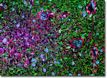
Glial fibrillary acidic protein (a type III intermediate filament protein), the calcium-binding protein calbindin, and heavy chain neurofilament subunits were immunofluorescently labeled in the coronal section of rat pons tissue presented above by treating the specimen with a cocktail of mouse anti-GFAP, rabbit anti-calbindin, and chicken anti-NF-H primary antibodies. The primary targets were visualized with goat anti-mouse, anti-rabbit, and anti-chicken secondary antibodies conjugated to Alexa Fluor 568, Alexa Fluor 647 (pseudocolored magenta), and Alexa Fluor 488, respectively. Hoechst 33342 was utilized to counterstain cell nuclei. Images were recorded with a 40x oil immersion objective using a zoom factor of 1.2 and sequential scanning with the 405-nanometer spectral line of a blue diode laser, the 488-nanometer spectral line of an argon-ion laser, the 543-nanometer line from a green helium-neon laser, and the 633-nanometer line of a red helium-neon laser. During the processing stage, individual image channels were pseudocolored with RGB values corresponding to each of the fluorophore emission spectral profiles unless otherwise noted above.
Contributing Authors
Nathan S. Claxton, Shannon H. Neaves, and Michael W. Davidson - National High Magnetic Field Laboratory, 1800 East Paul Dirac Dr., The Florida State University, Tallahassee, Florida, 32310.
