 |
 |
 |
|
||||||||||||||||||||||||
 | ||||||||||||||||||||||||
 | ||||||||||||||||||||||||
 | ||||||||||||||||||||||||
Confocal Microscopy Image Gallery
Rat Brain Tissue Sections
Cerebral Cortex
The cerebral cortex is the outer mantle of the brain that in humans is highly folded. Due to this folding, numerous fissures and crests (termed sulci and gyri) can be seen along the surface of the human brain. The cerebral hemispheres are usually each considered to be divided into six lobes (frontal, parietal, temporal, occipital, central, and limbic lobes) based upon the location of particular sulci and gyri, which vary little between individuals.

The cerebral cortices of other animals, however, do not necessarily have the same appearance as the human cortex. Rat brains, for example, exhibit almost entirely smooth cortices, and the brains of cats exhibit only a small number of convolutions compared to human brains. Despite massive differences in the extent of folding and total surface area, the thickness of the cerebral cortex remains nearly constant from species to species, differences being measurable in terms of only a few millimeters.
Neurons in the mammalian cerebral cortex display a laminar organization. The number of layers differs in certain cortical regions, but the vast majority of the cerebral cortex is composed of a minimum of six strata. This type of cortex is typically called neocortex, while more primitive cortical tissue with three layers is termed allocortex. The six primary layers of neocortex in order from innermost to outermost include the multiform layer, internal pyramidal layer, internal granular layer, external pyramidal layer, external granular layer, and the agranular (molecular) layer. Named for their predominant cell type, cortical layers vary in thickness and in neuron density.
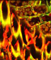
A coronal section of rat brain cerebral cortex (presented above) was immunofluorescently labeled for heavy chain neurofilament subunits with chicken anti-NF-H antibodies followed by goat anti-chicken secondary antibodies conjugated to Alexa Fluor 568 (red). Neurofilaments are specialized intermediate filaments chiefly found in the axons of neurons. In addition, the neuron-specific class III beta-tubulin isoform was targeted with mouse anti-beta-III-tubulin antibodies visualized with goat anti-mouse secondary antibodies conjugated to Alexa Fluor 488 (green). Nuclear DNA was counterstained with DRAQ5 (pseudocolored blue). Images were recorded with a 20x objective using a zoom factor of 2.0 and sequential scanning with the 488-nanometer spectral line of an argon-ion laser, the 543-nanometer line from a green helium-neon laser, and the 633-nanometer line of a red helium-neon laser. During the processing stage, individual image channels were pseudocolored with RGB values corresponding to each of the fluorophore emission spectral profiles unless otherwise noted above.
Additional Confocal Images of Rat Cerebral Cortex Tissue Sections
Targeting beta-III-Tubulin and Neurofilaments in Rat Brain Sections with Immunofluorescence - In a double immunofluorescence experiment, the coronal rat cerebral cortex section featured in this confocal image was fixed, permeabilized, blocked with 10-percent normal goat serum, and then treated with a cocktail of mouse anti-beta-III-tubulin (microtubules in neurons) and chicken anti-NF-H (neurofilaments) primary antibodies followed by goat anti-mouse and anti-chicken secondary antibodies (IgG) conjugated to Alexa Fluor 488 and Alexa Fluor 568, respectively. The specimen was counterstained for nuclei with DRAQ5, a far-red fluorescent DNA probe.
GFAP and NF-H Distribution in a Section of Rat Cerebral Cortex - Glial fibrillary acidic protein (GFAP) is a type III intermediate filament protein that is a primary structural element of the star-shaped cells known as astrocytes and neural stem cells. The protein was targeted in a coronal section of rat brain cerebral cortex by immunofluorescently labeling the specimen with primary rabbit anti-GFAP antibodies followed by goat anti-rabbit Fab fragments conjugated to Alexa Fluor 488. The section was also stained for neurofilaments with chicken anti-NF-H antibodies visualized with goat anti-chicken secondary antibodies conjugated to Alexa Fluor 568. Cell nuclei were labeled with DRAQ5.
Coronal Rat Brain Tissue Section Triple Stained for Calbindin, GFAP, and DNA - The calcium-binding protein calbindin and glial fibrillary acidic protein were immunofluorescently labeled in the thick section of rat cerebral cortex shown in this section by treating the specimen with a cocktail of rabbit anti-calbindin and mouse anti-GFAP primary antibodies followed by goat anti-rabbit and anti-mouse secondary antibodies conjugated to Alexa Fluor 488 and Alexa Fluor 568, respectively. DRAQ5, a DNA-interactive agent that exhibits preferential intercalation at AT base pairs, was utilized to target cell nuclei.
Neurofilaments and Glial Fibrillary Acidic Protein in the Rat Cerebral Cortex - A coronal section of rat brain cerebral cortex was immunofluorescently labeled for heavy chain neurofilament subunits with chicken anti-NF-H antibodies followed by goat anti-chicken secondary antibodies conjugated to Alexa Fluor 568. Neurofilaments, which are found primarily in the axons of neurons, are composed of light (NF-L) and medium (NF-M) chain subunits in addition to NF-H. Glial fibrillary acidic protein was also targeted in the tissue section by treating the specimen with rabbit anti-GFAP antibodies visualized with goat anti-rabbit secondary antibodies conjugated to Alexa Fluor 488. Nuclear DNA was counterstained with DRAQ5.
Contributing Authors
Nathan S. Claxton, Shannon H. Neaves, and Michael W. Davidson - National High Magnetic Field Laboratory, 1800 East Paul Dirac Dr., The Florida State University, Tallahassee, Florida, 32310.
