 |
 |
 |
|
||||||||||||||||||||||||
 | ||||||||||||||||||||||||
 | ||||||||||||||||||||||||
 | ||||||||||||||||||||||||
Confocal Microscopy Image Gallery
Plant Tissue Autofluorescence Gallery
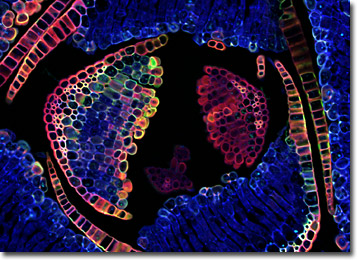
Hair-Cap Moss Leaves
Hair-cap mosses comprise the genus Polytrichum of the order Polytrichales. Both the genus and common name of these mosses make reference to the hairy calyptra characteristic of the sporophyte capsules (sporangia) before they reach maturity.

The capsules are very conspicuous, extending several inches into the air on long, narrow stalks and appearing similar to grains of wheat, causing some individuals to alternatively refer to the mosses as pigeon wheat. The calyptra forms from the wall of tissue that encases the archegonium of a female shoot present during the gametophyte stage of the moss life cycle.
Considerable masses of hair-cap mosses often naturally form in fields and peat bogs, and they are also sometimes cultivated as a ground cover or as garden subjects. The gametophyte generation of the mosses can be quite attractive. The most popular variety, P. commune, typically exhibits dark green, leathery leaf-like structures termed phyllids that may be held six inches or more in the air on erect stems. The phyllids are pointed and arranged spirally so that to an observer viewing them from above, the moss appears to be comprised of many small, leafy stars. Antheridia and archegonia grow at the tips of separate male and female plants, and the male reproductive organs form appealing flower-like structures on an annual basis.
Additional Confocal Images of Hair-Cap Moss Leaves
Hair-Cap Moss Leaves at High Magnification - Before the spores are released from the capsule of a hair-cap moss sporophyte, the operculum and calyptra are shed and the numerous peristomes around the mouth of the sporangium slowly uncurl. The spores are dispersed by the wind and germinate when they reach a suitable locale.
Contributing Authors
Nathan S. Claxton, Shannon H. Neaves, and Michael W. Davidson - National High Magnetic Field Laboratory, 1800 East Paul Dirac Dr., The Florida State University, Tallahassee, Florida, 32310.
