 |
 |
 |
|
||||||||||||||||||||||||
 | ||||||||||||||||||||||||
 | ||||||||||||||||||||||||
 | ||||||||||||||||||||||||
Confocal Microscopy Image Gallery
Mouse Hippocampal Neurons
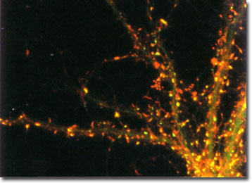
Hippocampal neurons expressing a green fluorescent protein (GFP) labeled postsynaptic density protein (green) were fixed and stained with rhodamine-phalloidin to visualize the location of cytoplasmic actin filaments (red) in the image presented below. In dendrites, the actin filaments are concentrated in the postsynaptic sites. The image was contributed by Shigeo Okabe from the Department of Anatomy and Cell Biology at the Tokyo Medical and Dental University in Japan.

The subcortical region of the brain known as the hippocampus is part of the limbic system. Horseshoe-shaped, the structure plays an important part in one’s emotions, sexuality, and spatial recognition capabilities. Most importantly, however, the hippocampus is actively involved in memory formation, and research has shown that damage to the area may result in the inability to remember people and events even if they have been recently encountered. Several scientific studies have indicated that this critical function of the hippocampus is closely connected to neurogenesis, which is the ability to grow new neurons. Though once considered to be a non-occurrence in adult vertebrates, an increasing body of evidence has indicated that neurogenesis continuously takes place in the hippocampi of healthy brains.
Confocal microscopy has been extremely important to the development of the modern conception of the hippocampus. By utilizing the technique in conjunction with various fluorescent markers, researchers have been able to acquire invaluable information regarding both morphological and functional aspects of this region of the brain. Some studies, for instance, have used confocal microscopy to obtain spatial and temporal resolution images of acute isolated hippocampal neurons, while others have differentiated their axonal and dendritic processes, as well as investigated their role in synaptic transmissions. Future developments in the field of neuroscience will also be readily facilitated by this extremely useful microscopy method, which enables three-dimensional visualization of images via optical sectioning and reconstruction.
