 |
 |
 |
|
||||||||||||||||||||||||
 | ||||||||||||||||||||||||
 | ||||||||||||||||||||||||
 | ||||||||||||||||||||||||
Confocal Microscopy Image Gallery
Human Skin Tissue
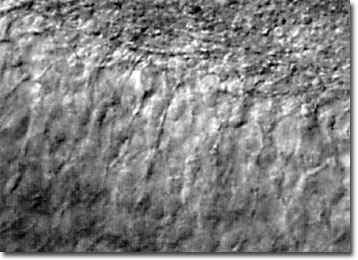
A thick section of living human skin tissue was imaged using infrared differential interference contrast (IR DIC) with a water immersion objective at a depth of approximately 80 micrometers to produce the image illustrated below. The specimen was illuminated with 750 nanometer light from a near infrared laser system. This striking image reveals the potential of laser scanning confocal microscopy when coupled with traditional contrast-enhancing techniques.

Skin, which is composed of two major layers, is the protective, flexible tissue that encloses the body of humans and other vertebrates. The epidermis is the skinís thin outer layer that primarily consists of closely packed cells, whereas the deeper, thicker dermis layer is chiefly composed of fibrous connective tissue that contains nerve endings, glands, and blood vessels. Together, the layers of skin act as a barrier against external influences, such as microbes and radiation, aid in maintaining proper moisture content inside the body, shield the internal organs against physical injury, and help sustain normal body temperature. Remarkably resilient, when it becomes damaged, the skin can often heal itself.
The confocal approach to microscopy has been perhaps the greatest advance in the study of skin tissues ever developed. By facilitating the examination of thick specimens and live cells, scientists have been able to obtain a more advanced understanding of the integumentary system than was ever before possible. The technique, which has generated a tremendous amount of interest among dermatologists and researchers around the world, has been widely utilized in a variety of applications. Among these are physiological studies, skin cancer research, monitoring of various integumentary therapies, live imaging of cutaneous vasculature, and in-vivo testing of cosmetic products.
