 |
 |
 |
|
||||||||||||||||||||||||
 | ||||||||||||||||||||||||
 | ||||||||||||||||||||||||
 | ||||||||||||||||||||||||
Confocal Microscopy Image Gallery
Drosophila Adult Brain
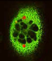
A three-dimensional volume rendering of confocal microscopy optical sections made from the brain of an adult fruit fly (Drosophila melanogaster) is presented in the image below. Mushroom bodies in the specimen were labeled with green fluorescent protein (GFP), and are highlighted in the green overlay. The image was contributed by Toshiro Aigaki in the Cytogenetics Department of the Tokyo Metropolitan University.

In their natural habitat, fruit flies feed upon the bacteria and yeast present in decaying and damaged fruits and vegetables, as implied by their common name. Due to their ability to invade economically important crops, such as strawberries and tomatoes, the tiny organisms are often considered agricultural pests. In the laboratory setting, however, Drosophila melanogaster is seen in quite a different light. Simple to breed and maintain, the ubiquitous fruit fly is viewed as an invaluable experimental specimen. For more than 50 years, it has been commonly utilized in cellular and molecular genetics research and, more recently, has become a cornerstone species in developmental biology studies.
Mushroom bodies are vital centers for high-order sensory assimilation and learning in Drosophila melanogaster and other insects. The modern understanding of these bodies has been greatly enhanced through the utilization of confocal microscopy techniques, especially optical sectioning, fluorescent marking, and three-dimensional visualization. Such methods have also yielded a tremendous amount of other significant anatomical, biological, and physiological information about the fruit fly. Moreover, these findings have been combined to create critical scientific resources for further research, such as three-dimensional maps of antennal lobes and other minute structures, highly detailed neuroanatomical descriptions of the central and peripheral nervous system, and interactive virtual dissection programs that would not have been possible without the use of confocal instrumentation.
