 |
 |
 |
|
||||||||||||||||||||||||
 | ||||||||||||||||||||||||
 | ||||||||||||||||||||||||
 | ||||||||||||||||||||||||
Confocal Microscopy Image Gallery
Normal African Green Monkey Kidney Epithelial Cells (Vero Line)
Y. Yasumura and Y. Kawakita at the Chiba University in Chiba, Japan established the Vero epithelial cell line in the early 1960s from the kidney tissue of an adult African green monkey. The cells are a standard cell line utilized in many laboratories, especially for vaccine production, transfections, and the detection of verotoxins.

The Vero cell line is negative for reverse transcriptase, which indicates an absence of integral retrovirus genomes, and has been demonstrated to be resistant to a variety of viruses, including Apeu, Nepuyo, Caraparu, Stratford, Madrid, and Ossa viruses. The cells have been shown to be susceptible to an even larger number of viruses than to which they are resistant. Simian viruses 5 and 40, polioviruses, rubeola, rubellavirus, reoviruses, simian adenoviruses, Paramaribo, Getah, Ndumu, Pixuna, Guaroa, Murutucu, Germiston, Ross River, Semliki Forest, Kokobera, Modoc, Pongola, and Tacaribe are among the many viruses to which the Vero line is known to exhibit susceptibility.
Some scientists are currently carrying out investigations with Vero cells that could eventually lead to the development of innovative new treatments for diabetes. Diabetes is one of the leading causes of kidney disease and is considered to result from two different causes. Related to the inability to metabolize glucose, diabetes can occur due to damage to the pancreatic beta cells that usually produce insulin (Type 1) or can be associated with a diminished bodily response to insulin that is produced (Type 2). Recently, a team of scientists in Ireland working with Vero cells found that the cells exhibit promise for use in a type of cell therapy that potentially could aid individuals with Type 1 diabetes. As posited by the research team, Vero cells could be modified to act as substitutes for damaged beta cells. The benefit of such an approach, as opposed to utilizing real beta cells from donors, would be that the Vero-based beta cell substitutes could potentially go unrecognized by the body’s autoimmune system, which is responsible for attacking and destroying the insulin-producing cells in patients with the Type 1 disease.
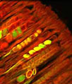
Filamentous actin was targeted in the culture of African green monkey kidney cells presented in the digital image above with BODIPY FL conjugated to phallacidin (pseudocolored blue), a bicyclic peptide derived from a toxic mushroom. In addition, the cells were treated with the mitochondrion-selective stain MitoTracker Red CMXRos (pseudocolored yellow) and a far-red fluorescent DNA probe, DRAQ5. Images were recorded with a 60x oil immersion objective using a zoom factor of 2.0 and sequential scanning with the 488-nanometer spectral line of an argon-ion laser, the 543-nanometer line from a green helium-neon laser, and the 633-nanometer line of a red helium-neon laser. During the processing stage, individual image channels were pseudocolored with RGB values corresponding to each of the fluorophore emission spectral profiles unless otherwise noted above.
Additional Confocal Images of African Green Monkey Kidney Epithelial (Vero) Cells
African Green Monkey Kidney Cells Stained with MitoTracker Red CMXRos, Alexa Fluor 488, and TO-PRO-3 - The mitochondrial network in the culture of Vero epithelial cells presented in this section was labeled with MitoTracker Red CMXRos (pseudocolored yellow), a derivative of X-rosamine. In addition, the cells were stained with Alexa Fluor 488 conjugated to phalloidin and TO-PRO-3, targeting filamentous actin and nuclear DNA, respectively.
Distribution of Mitochondria and F-Actin in Vero Cell Cultures - The adherent monolayer African green monkey kidney cell culture presented in this section was labeled for the cytoskeletal filamentous actin and intracellular mitochondrial networks with Alexa Fluor 488 conjugated to phalloidin and MitoTracker Red CMXRos, respectively. Nuclei present in the epithelial cells were counterstained with a carbocyanine monomer, TO-PRO-3 (pseudocolored cyan).
Examining the Proximity Between the Nucleus and the Mitochondrial Network in Monkey Kidney Epithelial Cells - A log phase monolayer culture of Vero cells was treated with MitoTracker Red CMXRos (pseudocolored yellow) in growth medium for one hour, washed, and fixed with 3.7-percent paraformaldehyde in medium containing serum. After washing and permeabilization, the cells were blocked with bovine serum albumen in PBS and labeled with BODIPY FL conjugated to phallacidin. The nuclei were subsequently counterstained with DRAQ5, an anthraquinone that exhibits preferential intercalation at AT base pairs.
Contributing Authors
Nathan S. Claxton, Shannon H. Neaves, and Michael W. Davidson - National High Magnetic Field Laboratory, 1800 East Paul Dirac Dr., The Florida State University, Tallahassee, Florida, 32310.
