 |
 |
 |
|
||||||||||||||||||||||||
 | ||||||||||||||||||||||||
 | ||||||||||||||||||||||||
 | ||||||||||||||||||||||||
Confocal Microscopy Image Gallery
Horse Dermal Fibroblast Cells (NBL-6 Line)
Initiated from the dermal tissue of a 4-year-old female horse (Equus caballus) of the quarterhorse strain, the NBL-6 cell line (also called the E. Derm line) has played an important role in equine viral arteritis (EVA) research. EVA is a contagious respiratory disease caused by a small, enveloped RNA virus.

The fibroblast cell line is also commonly utilized to propagate viruses for equine vaccine production. NBL-6 cells are known to be susceptible to reovirus 3, herpes simplex, vesicular stomatitis (Ogden strain), and vaccinia and are resistant to coxsackieviruses A9 and B5, poliovirus 2, and adenovirus 5. Senescence of the fibroblasts usually takes place after about 40 passages of the cells.
Dermal tissue, such as that utilized as the initial source of the NBL-6 line, is an integumentary layer of connective tissue located directly beneath the epidermis. The dermis is derived from the mesoderm and is chiefly composed of fibroblasts, but the stratum contains macrophages, mast cells, and various other connective tissue cells as well. Embedded within the dermis are a number of important structures, including blood vessels, sweat glands, sebaceous glands, lymph channels, nerve endings, and hair follicles. Large amounts of water are usually held by glycosaminoglycans present in the dermis, which keeps the skin turgid. Damage to the dermis is much more serious than damage to the epidermis, with burns affecting the layer being classified as either second- or third-degree depending on the depth of tissue affected.
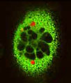
The actin cytoskeleton was targeted in the culture of NBL-6 cells presented with Alexa Fluor 488 conjugated to phalloidin, a bicyclic peptide derived from a toxic mushroom. In addition, the cells were treated with the mitochondrion-selective stain MitoTracker Red CMXRos and a far-red fluorescent DNA probe, DRAQ5 (pseudocolored cyan). Images were recorded with a 60x oil immersion objective using a zoom factor of 2.5 and sequential scanning with the 488-nanometer spectral line of an argon-ion laser, the 543-nanometer line from a green helium-neon laser, and the 633-nanometer line of a red helium-neon laser. During the processing stage, individual image channels were pseudocolored with RGB values corresponding to each of the fluorophore emission spectral profiles unless otherwise noted above.
Additional Confocal Images of Horse Dermal Fibroblast Cells (NBL-6) Cells
Horse Dermal Fibroblasts Triple Labeled with BODIPY FL, MitoTracker Orange CMTMRos, and TO-PRO-3 - The distribution of filamentous actin and mitochondria in a culture of horse dermal fibroblast cells was visualized with BODIPY FL conjugated to phallacidin and MitoTracker Orange CMTMRos (pseudocolored yellow). Cell nuclei were counterstained with TO-PRO-3, a carbocyanine monomer with long-wavelength red fluorescence.
Distribution of F-Actin and Mitochondria in NBL-6 Cells - A triple fluorophore combination of MitoTracker Red CMXRos, Alexa Fluor 488 conjugated to phalloidin, and DRAQ 5 (pseudocolored cyan) was used to label an adherent log phase culture of NBL-6 cells for mitochondria, the filamentous actin network, and nuclear DNA, respectively. The cells were first treated with the MitoTracker probe in growth medium for one hour, washed and fixed with paraformaldehyde (prepared in growth medium), permeabilized, and blocked with bovine serum albumen.
Contributing Authors
Nathan S. Claxton, Shannon H. Neaves, and Michael W. Davidson - National High Magnetic Field Laboratory, 1800 East Paul Dirac Dr., The Florida State University, Tallahassee, Florida, 32310.
