 |
 |
 |
|
||||||||||||||||||||||||
 | ||||||||||||||||||||||||
 | ||||||||||||||||||||||||
 | ||||||||||||||||||||||||
Confocal Microscopy Literature
Recommended Books on Confocal Microscopy
A surprisingly limited number of books dealing with various aspects of laser scanning and spinning disk confocal microscopy and related techniques are currently available from the booksellers. This section lists the FluoView Resource Center website development team's top 12 recommended books. Although the volumes listed in this section deal pricipally with confocal microscopy and related methodology, there exist a number of additional books that contain focused treatments of the materials described below, and these should also be consulted for specific techniques and timely review articles.
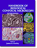
Handbook of Biological Confocal Microscopy - James B. Pawley (editor). With numerous contributions from leading researchers in the field, this second edition is a compendium of excellent review articles covering virtually all of the important aspects of laser scanning confocal microscopy. Included are reviews of light sources (both coherent and incoherent), objectives, optical systems, detectors, image processing, optical sectioning, deconvolution, reflection confocal, fluorophores, image contrast, specimen preservation, imaging living cells, multiphoton, lifetime imaging, and real time imaging. An extensive bibliography is also provided, although it is now somewhat dated. The appendices of the text contain useful diagrams of the scanning heads offered by major commercial manufacturers, and several articles contain information about the fundamental aspects of resolution and imaging in optical microscopy. A new volume is in the works, but this edition (published in 1995) is still available and contains one of the best collections of review articles available in a single location.
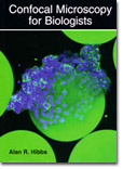
Confocal Microscopy for Biologists - Alan R. Hibbs. Written as a guide for biological scientists, this 467-page treatise proves to be an excellent introduction to both the theory and techniques of confocal microscopy. The foundation is prepared with a chapter on the basic concepts surrounding optical phenomena in microscopy, aberrations, imaging modes, light sources, and a discussion of the main components comprising modern research microscopes. Next, the book discusses confocal microscopy hardware, image collection, digital imaging and software available for image processing, as well as how to prepare images for publication in print media or on the web. A review of fluorescence, synthetic probes, and basic immuno-labeling techniques for cells and tissues is targeted at optimizing staining and visualization of specimens for confocal microscopy. Live-cell imaging is covered in detail with a thorough discussion of enviromental chambers, cell nutrient and pH requirements, and suggestions for imaging strategies. The book also reviews Internet sites dealing with confocal microscopy and lists technical supplies (and their sources) necessary for successful imaging. Approximately 100 pages of appendicies review manufacturer-specific information about commercial instruments. This book is highly recommended for beginners and advanced researchers alike.
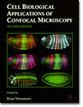
Cell Biological Applications of Confocal Microscopy - Brian Matsumoto (editor). Published as Volume 70 in the series Methods in Cell Biology, this second edition is a collection of review articles by experts in the field of confocal microscopy and live-cell imaging. Included is an broad introduction to laser scanning techniques and instruments, a discussion of scanning-disk instruments, and a summary of multiphoton imaging in the biological sciences. Other subjects address multicolor immunofluorescence, objective selection, imaging considerations, and suggestions on establishing and operating a core facility. Several chapters are dedicated to specific specimens, such as Tetrahymena, Drosophila, and amphibian embryos, while others focus on ratiometric dyes, calcium indicators, and pH-sensitive probes. The highly specific nature of several reviews will limit their interest to a narrow spectrum of readers, but many of the chapters have appeal to a wide audience.
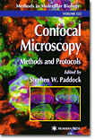
Confocal Microscopy: Methods and Protocols - Stephen W. Paddock (editor). Published as Volume 122 of the popular Methods in Molecular Biology series, the book represents an excellent compendium of the current methodology and techniques employed for specimen preparation, imaging, measurements, and data processing in confocal microscopy. Along with an introductory chapter on the basic aspects of confocal microscopy, the book contains a very nice treatment of the practical considerations necessary for successful confocal imaging. A number of detailed specific protocols for confocal imaging are included: gene expression with antibody probes, fluorescence in situ hybridization (FISH) experiments, plants, yeast, sea urchin fertilization, oocytes and embryos, Zebrafish, fluorescent proteins, calcium indicators, pH measurement, thick tissues, and cell volume. Later chapters review the presentation of confocal images, preparation of three-dimensional illustrations, digital video production, and information management. The wide variety of protocols outlined in this book make it an essential volume for the library of any serious confocal microsopist.
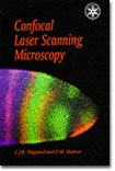
Confocal Laser Scanning Microscopy - Colin J. R. Sheppard and David M. Shotton. Almost considered as a "pocket guide" to confocal microscopy, this volume is published in the Royal Microscopical Society Microscopy Handbooks series (Volume 38). The 106-page book presents a very nicely crafted overview of basic concepts in confocal microscopy, including scanning modes, image formation, photodetectors, objectives, and microscope alignment. A well-written chapter entitled "Performance of Confocal Microscopes" reviews optical sectioning, the point spread function, axial and lateral resolution, sensitivity, dynamic range, and geometric linearity. Later chapters discuss image processing, confocal fluorescence, biological applications, industrial applications, and specialized techniques such as near-field, 4pi, theta, two-photon, and other non-linear confocal techniques. This book is a very good introduction to confocal microscopy and is recommended for both beginners and advanced students.
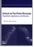
Confocal and Two-Photon Microscopy: Foundations, Applications, and Advances - Alberto Diaspro (editor). Containing 26 chapters written by recognized experts in the field of confocal and multiphoton microscopy, this 567-page book addresses both theoretical concepts and applied research. Included are chapters on basic principles, fluorophores, resolution, contrast, aperture restrictions, aberrations, fiber optic couplers, sampling, and image processing. Other chapters review live cell imaging, neuronal growth and morphology, tissue physiology, plant biology, spectroscopy, calcium imaging, photobleaching, superresolution, three-dimensional imaging, and characterization of integrated circuits by two-photon microscopy. Because so many of the chapters represent authoritive review articles with extensive reference lists, this volume will be essential reading for researchers in a wide spectrum of disciplines.

Confocal Microscopy (Methods in Enzymology - Volume 307) - P. Michael Conn (editor). This 663-page book documents many diverse applications for confocal microscopy in disciplines that broadly span the biological sciences. Many of the methods presented include shortcuts and conveniences that are not usually incorporated into general review articles, and the techniques are described in a context that allows comparisons to other related methodologies. Included are sections on theoretical and practical considerations, general techniques, measurement of subcellular relations and volume determinations, imaging of processes (such as phagocytosis, secretion, receptor-ligand binding, etc.), ion imaging, applications for specialized tissues, and the imaging of viruses and fungi. Although many topics are not covered in this volume, the treatment provides a substantial and current overview of the extant methodology in the field and a view of its rapid development.
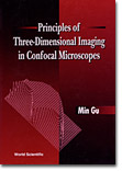
Principles of Three-Dimensional Imaging in Confocal Microscopes - Min Gu. This volume discusses a variety of principles in laser scanning confocal microscopy in a unique manner based on the concept of the three-dimensional transfer functions developed by the author and his colleagues over the past few years. Using the transfer functions, resolving power in three-dimensional confocal imaging can be defined in a unified manner and different optical configurations can be compared. In addition, optical section images of thick specimens can be modeled with an insight to their interrelationship. The book employs advanced mathematical treatments to describe three-dimensional Fourier optical principles, point spread functions, and transfer functions. Chapters include brightfield microscopy with a finite-sized detector, confocal fluorescence, two-photon, fiber optics, short pulse laser microscopy, high numerical aperture objectives, and the significance of three-dimensional transfer functions.
Three-Dimensional Confocal Microscopy: Volume Investigation of Biological Systems - John K. Stevens, Linda R. Mills, and Judy E. Trogadis (editors). The integration of laser scanning and spinning disk confocal microscopy techniques with volume investigations has led to an unprecented ability to examine spatial relationships between cellular structure and function. The goal of this volume is to familiarize the reader with these technologies and to demonstrate their applicability to a wide range of biological and clinical problems. Chapter topics include noise sources, fluorescent probes, display methods, volume reconstruction, and a host of biological applications. The final section addresses alternative approaches and future applications. Even though the book was published in the mid-1990s, many of the techniques described by the expert authors apply to current confocal microscopy investigations.
Confocal Scanning Optical Microscopy and Related Imaging Systems - Timothy R. Corle and Gordon S. Kino. Providing a comprehensive introduction to the field of scanning confocal microscopy, this volume concentrates on laser instruments, the optical interference microscope, and provides a discussion of the theory and design of near-field systems. The text discusses the practical aspects of building a confocal scanning optical microscope or optical interference microscope, and the applications of these microscopes to phase imaging, biological imaging, and semiconductor inspection. A thorough theoretical discussion of the axial and transverse resolution is given with emphasis placed on the practical results of calculations and how these can be applied to the understanding and operation of these complex microscopes. Additional topics include spinning disk and slit confocal microscopes, vector field theory for resolution, and film thickness measurements.
Confocal Microscopy - Tony Wilson (editor). Although out of print and somewhat difficult to obtain, this volume reviews the state of confocal imaging in the late 1980s and early 1990s. Many of the invited articles contain information that is of current interest to researchers and students. Included are reviews on software, electronics, optical sectioning, three-dimensional imaging, optical systems, pinhole apertures, fluorescence, brightfield techniques, spinning disk techniques, measurements, and treatments on biological applications. The final chapters include a discussion of semiconductor metrology, real-time scanning optical microscopes, and confocal interference microscopy.
Selected Papers on Confocal Microscopy - Barry R. Masters and Brian J. Thompson (editors). Published as Volume 131 of the International Society for Optical Engineering (SPIE) Milestone Series, this book contains numerous reprints of original papers in confocal microscopy and related fields. Section headings include: surveys of early history, early developments in confocal microscopy, the role of detector geometry, superresolution, fluorescence imaging, interference and differential confocal microscopy, three-dimensional imaging, slit scanning instruments, new directions in biological and medical research, optical sectioning, 4pi, a bibliography, and selected patents. The volume is an excellent source of older original material, which is often difficult to obtain.
