Specimen Preparation Using Synthetic Fluorophores and Immunofluorescence
Rat Diaphragm Smooth Muscle Tissue

|
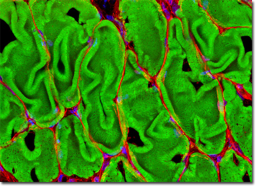
Presented above is a laser scanning confocal image revealing the extensive filamentous actin network present in the smooth muscle tissue of a thin (8-micrometer) cryosection of rat diaphragm. The tissue cryosection was labeled with a cocktail containing Alexa Fluor 488 conjugated to phalloidin (staining actin) and Texas Red-X conjugated to wheat germ agglutinin (targeting lectins). In addition, nuclei in the specimen were counterstained with Hoechst 33342. Images were recorded in grayscale with an Olympus FluoView FV1000 coupled to a BX-81 inverted microscope using Argon-ion (488 nanometer line), violet diode (405 nanometers), and green helium-neon (543 nanometers) lasers. During the processing stage, individual image channels were pseudocolored with RGB values corresponding to each of the fluorophore emission spectral profiles. View a larger version of this digital image. |
 |
 |
 |
 |
 |
 |
 |